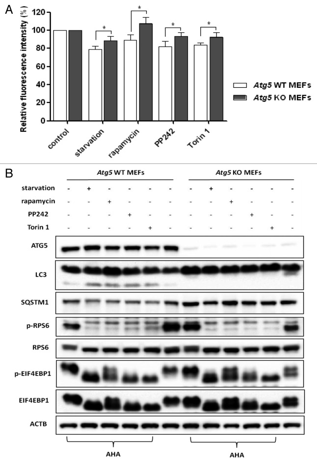
Figure 4. Defective autophagy impaired long-lived protein degradation. (A) Atg5 WT and KO MEFs were labeled with AHA and then treated with starvation or MTOR inhibitors for 3 h, as described in Figure 2A. Data for the relative signal intensity were expressed as the ratio of treated cells to control cells, as mean ± SD from 3 independent experiments, *P < 0.05, the Student t test. (B) Atg5 WT and KO MEFs were treated as described in (A), harvested and proteins from cell lysates were analyzed by western blot. ACTB served as the loading control.

An official website of the United States government
Here's how you know
Official websites use .gov
A
.gov website belongs to an official
government organization in the United States.
Secure .gov websites use HTTPS
A lock (
) or https:// means you've safely
connected to the .gov website. Share sensitive
information only on official, secure websites.
