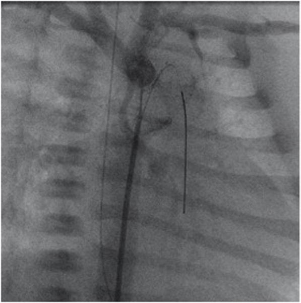
Figure 3.

Right anterior oblique angiography performed through a 4F multipurpose catheter positioned at the junction of the arterial duct (stented by an open cell self-expanding SinusSuperflex-DS stent). The extreme hypoplastic ascending aorta is connected to a rather well developed aortic arch. A coronary soft-tip wire is seen passing retrograde through the ductal stent into the right ventricular outflow tract.
