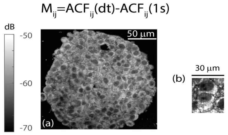
Fig. 7.

Dynamic contrast with FFOCT provided by the fast decay of the ACF, Mij. (a) Spheroid imaged at the 60 μm imaging depth of this study with the 0.8 numerical aperture FFOCT prototype. (b) Individual cells imaged with the prototype (linear gray map with 2 dynamic range).
