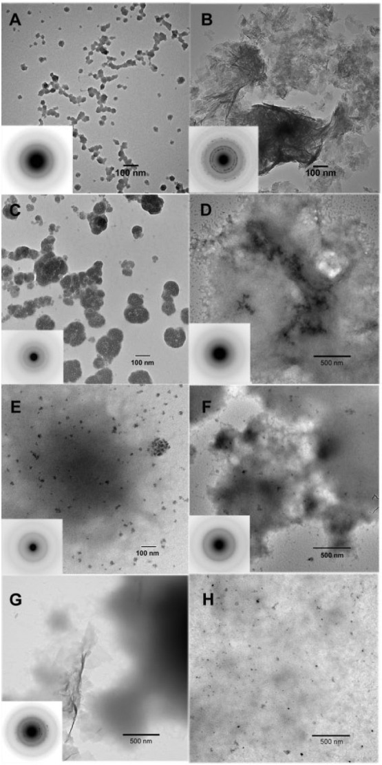
Figure 1.

Transmission electron microscopy (TEM) and selected area electron diffraction (SAED) (insets) analyses of calcium phosphate mineral products formed. As a control, in the absence of protein, amorphous calcium phosphate (ACP) particles were predominantly observed at 24 h (A). Subsequently, these initially formed ACP nanoparticles were found to almost completely transform into large agglomerations of randomly arranged plate-like hydroxyapatite (HA) crystals at 48 h (B). In the presence of 1 mg/mL P173 alone, only ACP particles were observed at 24 h (C) and 48 h (D). In the presence of 1 mg/mL P148 (E–G) without MMP20, ACP was observed at both 24 h (E) and 48 h (F), although some randomly disordered plate-like HA crystals were also found at 48 h (G). In the presence of 2 mg/mL P148 without MMP20, however, ACP particles were almost exclusively found at 48 h (H), along with protein aggregates that tended to exhibit greater contrast.
