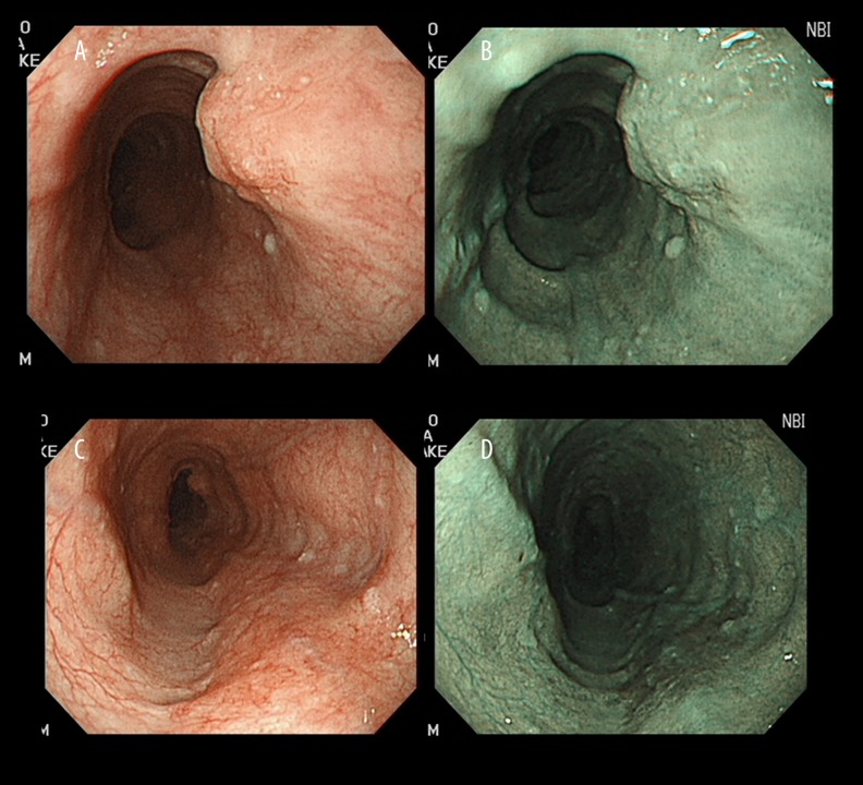
Figure 1.
(A) Case 4. Conventional endoscopy shows a protruding lesion (0–Is) located 37 cm from the incisors (lesion 1). (B) There are few brownish areas at the lesion on narrow-band imaging. (C) Case 4. Conventional endoscopy shows a flat lesion (0–IIb) located 30 cm from the incisors. (D) There is a brownish area at the lesion on narrow-band imaging (lesion 2).

