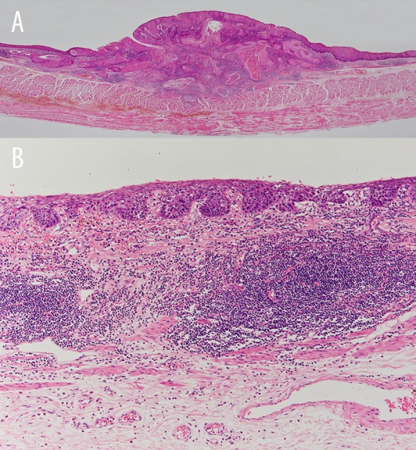
Figure 2.

(A) Case 4. Microscopic findings. Tumor depth was T2, ly0, v0 for lesion (lesion 1). (B) T1a-LPM, ly0, v0 for lesion 2 (lesion 2). Epithelial thickening and increased vascularity in the mucosal or submucosal layers were seen at both lesions.
