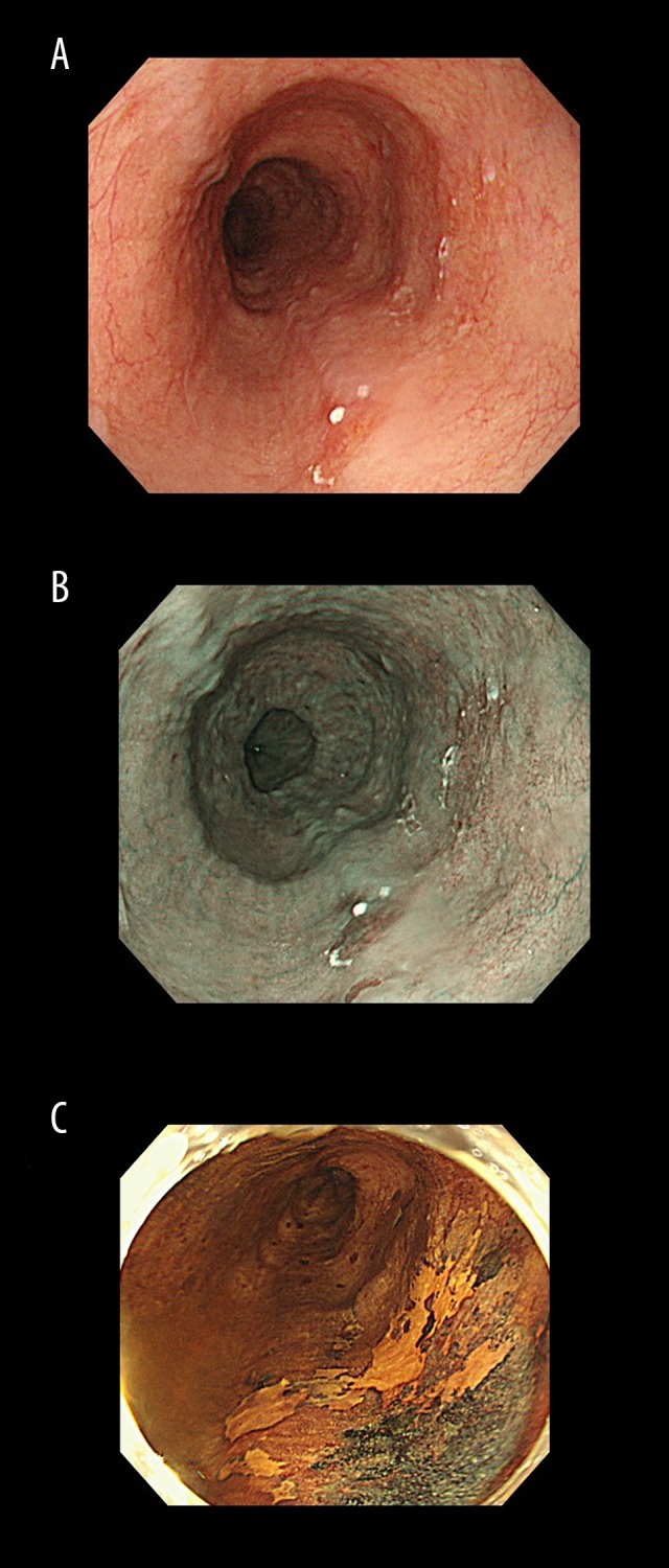
Figure 3.

(A) Case 6. Endoscopic findings. Conventional endoscopy shows slightly depressed lesion (0-IIc) at 29–30 cm from the incisor teeth. (B) Case 6. There is a brownish area at the lesion on narrow-band imaging. (C) Case 6. Chromoendoscopy reveals that the lesion is not stained by iodine.
