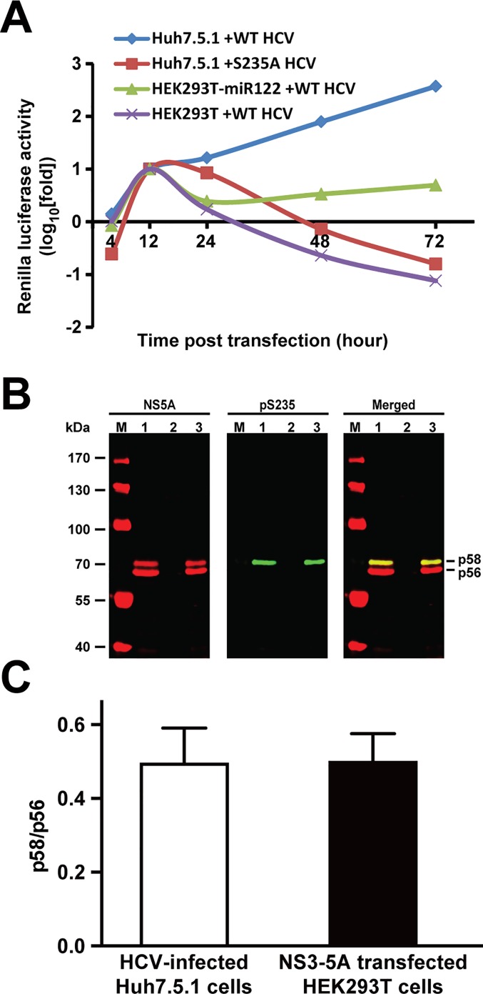
Fig 1. The HEK293T kidney cells recapitulated HCV NS5A phosphorylation and functions as seen in the HCV-infected Huh7.5.1 liver cells.
(A) Reporter virus assay. The HEK293T cells were transfected with the wild type (WT) full length reporter HCV genomic RNA (5’C19Rluc2AUbi) with or without the miR-122 expression vector before the measurements of the Renilla luciferase activity. The Huh7.5.1 cells transfected with the full length WT or S235A replication-defective reporter HCV genomic RNA serves as controls. (B) Representative and (C) summary of the immunoblotting for NS5A and NS5A phosphorylation at serine 235 (pS235) in the HCV (J6/JFH1)-infected Huh7.5.1 cells (7 days after infection, lane 1) and in the HEK293T cells transfected without (lane 2) or with the NS3-5A expression construct (2 days after transfection, lane 3). Lane M indicates protein size markers. Hypo- and hyper-phosphorylated NS5A are labeled as p56 and p58, respectively. Protein abundance was measured with the Li-Cor Odyssey scanner and software. Values are Mean ± SEM (n = 3).

