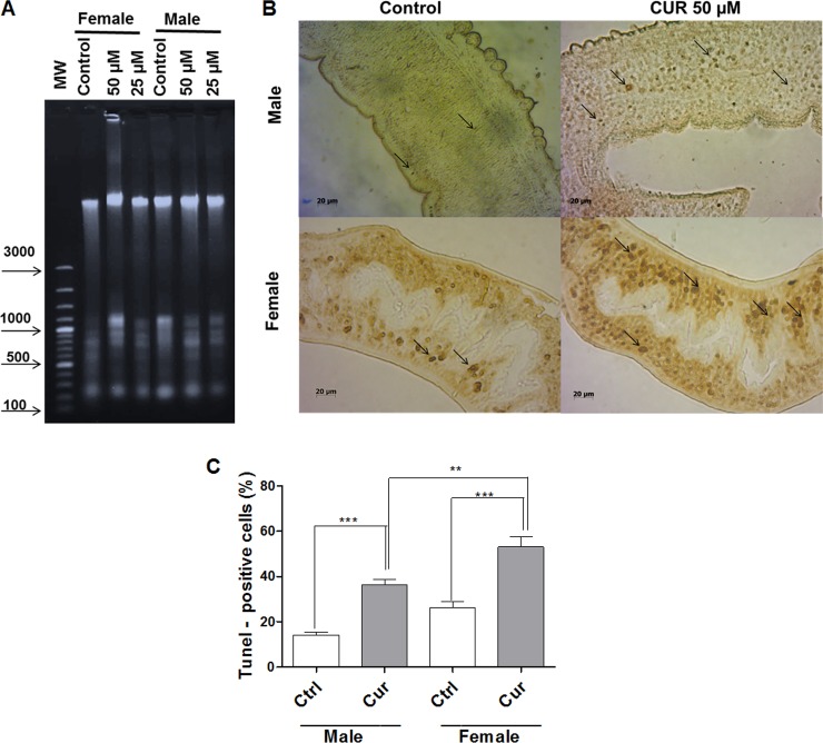
Fig 4. CUR induces DNA fragmentation and damage in adult S. mansoni worms.
Couples of adult worms were incubated with CUR at the indicated concentrations for 24 h. After incubation, female and male S. mansoni worms were separated and analyzed. In the negative control groups, couples of adult worms were incubated with RPMI 1640 medium with 0.1% DMSO. (A) Genomic DNA of adult female and male worms was extracted as described in material and methods, and 600 ng of the DNA was run in 2% agarose gel containing 1% GelRed (1:500) (MW Molecular weight marker). The experiments were repeated twice, and ten couples of adult worms were evaluated in each experiment. (B) TUNEL-stained light micrographs of adult female and male worm sections (arrows indicate the dark brown-stained apoptotic nuclei). (C) Histograms indicate the percentage of TUNEL-positive cells. For each experiment, at least 100 cells were analyzed. Values are expressed as the mean ± S.E.M of three independent experiments. An asterisk indicates statistically significant differences as compared to the negative control group (RPMI 1640 medium with 0.1% DMSO) or when male and female worms were compared (**p < 0.01, ***p < 0.001).

