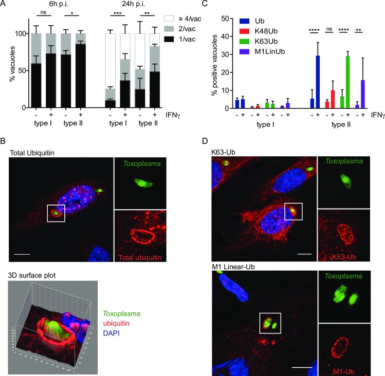
Fig 1. IFNγ-mediated K63-linked ubiquitination of type II Toxoplasma vacuoles.
(A) Replication of both type I and type II Toxoplasma is diminished in IFNγ-stimulated HUVEC at 6h and more substantially at 24h post invasion. HUVEC were infected (-/+ stimulation with 50 units/ml IFNγ for 18h) with type I or type II parasites for 6h and 24h. Results from ≥3 experiments are shown. Significance was calculated using 2way ANOVA, *, p≤0.05, **, p≤0.01, ***, p≤0.001 and ns, not significant. (B) Ubiquitin is recruited to the PV of type II Toxoplasma in HUVEC. HUVEC were stimulated with 50 units/ml IFNγ for 18h before infecting with type II Toxoplasma for 2.5h. Confocal image of a representative vacuole is shown. Scale bar 10μm. The ubiquitin staining is mainly at the PV and not substantially on the parasite. 3D surface profile (Image J) of the pixel intensity over a ubiquitin-stained vacuole containing type II Toxoplasma, on a confocal section. Ubiquitin intensity (red) and parasite (green) are shown. (C) Ubiquitin around the type II Toxoplasma vacuole is mostly of the K63 linkage. Antibodies specific for ubiquitin linkages K63, K48 and M1 linear were used alongside an antibody staining total ubiquitin. Quantitation of the indicated linkage-specific ubiquitin-positive PVs, at 2.5h p.i., is shown. The mean of 3 experiments is shown. Significance was calculated using 2way ANOVA, **, p≤0.01, ****, p≤ 0.0001 and ns, not significant. (D) K63-linked and M1-linear ubiquitin accumulated at the type II Toxoplasma PV. Confocal images were taken of HUVEC stimulated with 50 units/ml IFNγ for 18h, before infecting with type II Toxoplasma for 2.5h. Infected cells were then fixed and stained with linkage-specific ubiquitin antibodies. Representative images are shown. Scale bar 10μm.

