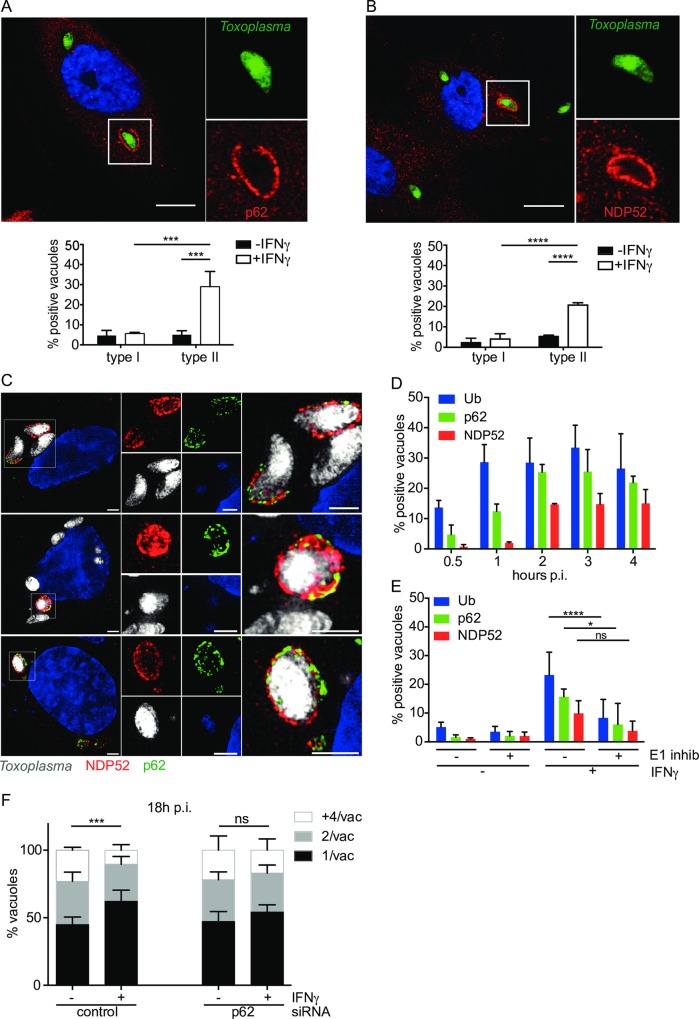
Fig 2. p62 and NDP52 are recruited to microdomains on the type II Toxoplasma PV in dependence of ubiquitination.
(A) p62 accumulates specifically at the type II Toxoplasma PV in IFNγ-stimulated HUVEC. Confocal images were taken of IFNγ-stimulated HUVEC infected with type II Toxoplasma for 2.5h, fixed and stained with p62 antibody and a representative image is shown. Scale bar 10μm. Quantitation of p62-positive PVs under the indicated conditions is shown, 2.5h p.i. The mean of 3 experiments is shown. Significance was calculated using 2way ANOVA, ***, p ≤ 0.001. (B) NDP52 accumulates specifically at the type II Toxoplasma PV in IFNγ-stimulated HUVEC. Confocal images were taken of HUVEC infected with type II Toxoplasma for 2.5h, fixed and stained with NDP52 antibody and a representative image is shown. Scale bar 10μm. Quantitation of NDP52-positive PVs under the indicated conditions is shown, 2.5h p.i.. The mean of 3 experiments is shown. Significance was calculated using 2way ANOVA, ****, p≤ 0.0001. (C) p62 and NDP52 occupy overlapping microdomains at the type II Toxoplasma PV in IFNγ-stimulated HUVEC. Superresolution Structured Illumination Microscopy images of 3 representative vacuoles demonstrated mostly a patchy and distinct localisation of p62 and NDP52 with some small overlapping microdomains. Scale bar 2μm. (D) Host defence proteins p62 and NDP52 are recruited to the PV subsequent to ubiquitin binding to the PV. Ubiquitin accumulates on the type II PV within 30min-1h p.i., reaching a maximum already at 1h. p62 sequentially follows the ubiquitin recruitment, with a maximum at 2–4h p.i. and NDP52, appearing last to maximum levels at 2h p.i and is maintained until at least 4h p.i. Quantitation of recruitment-positive type II PVs, at the indicated time-points p.i. is shown. The mean of ≥3 experiments is shown. (E) p62 and NDP52 recruitment to the PV depends upon the ubiquitination of the PV. Inhibition of ubiquitination in HUVEC with the E1 inhibitor UBEI-41 leads to a reduction in ubiquitin as well as p62 and NDP52 at the vacuole of type II Toxoplasma. IFNγ-stimulated HUVEC were pre-incubated with 50μM UBEI-41 for 2h before washing and infecting with type II Toxoplasma for 2.5h. Cells were stained for ubiquitin, p62, NDP52 and positive staining recorded. The mean of ≥3 experiments is shown. Significance was calculated using 2way ANOVA, *, p ≤ 0.05, ****, p ≤ 0.0001 and ns, not significant. (F) Knockdown of p62 by siRNA leads to the loss of IFNγ-restriction of type II Toxoplasma. siRNA nucleofection of p62 was used to knock down the protein in HUVEC, comparing to a control siRNA. 24h after siRNA nucleofection, the cells were stimulated with 50units/ml IFNγ for a further 24h. Infection with type II Toxoplasma was then allowed to proceed for 18h and the effect on replication recorded in fixed cells by scoring the numbers of parasites/vacuole in >100 vacuoles. Significance was determined by 2way ANOVA, ***, p ≤ 0.001 and ns, not significant.

