
Figure 4. Hematoxylin & eosin staining of control, mild and moderate FR mouse placentas at 4× magnification.
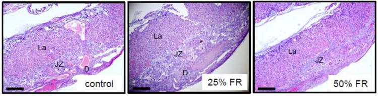
Representative murine placental sections stained by H&E from control, mild- and moderate-FR exposed mice (n=8/group). La: labyrinth; JZ: junctional zone; D: decidua. Scale bars represent 500μm.