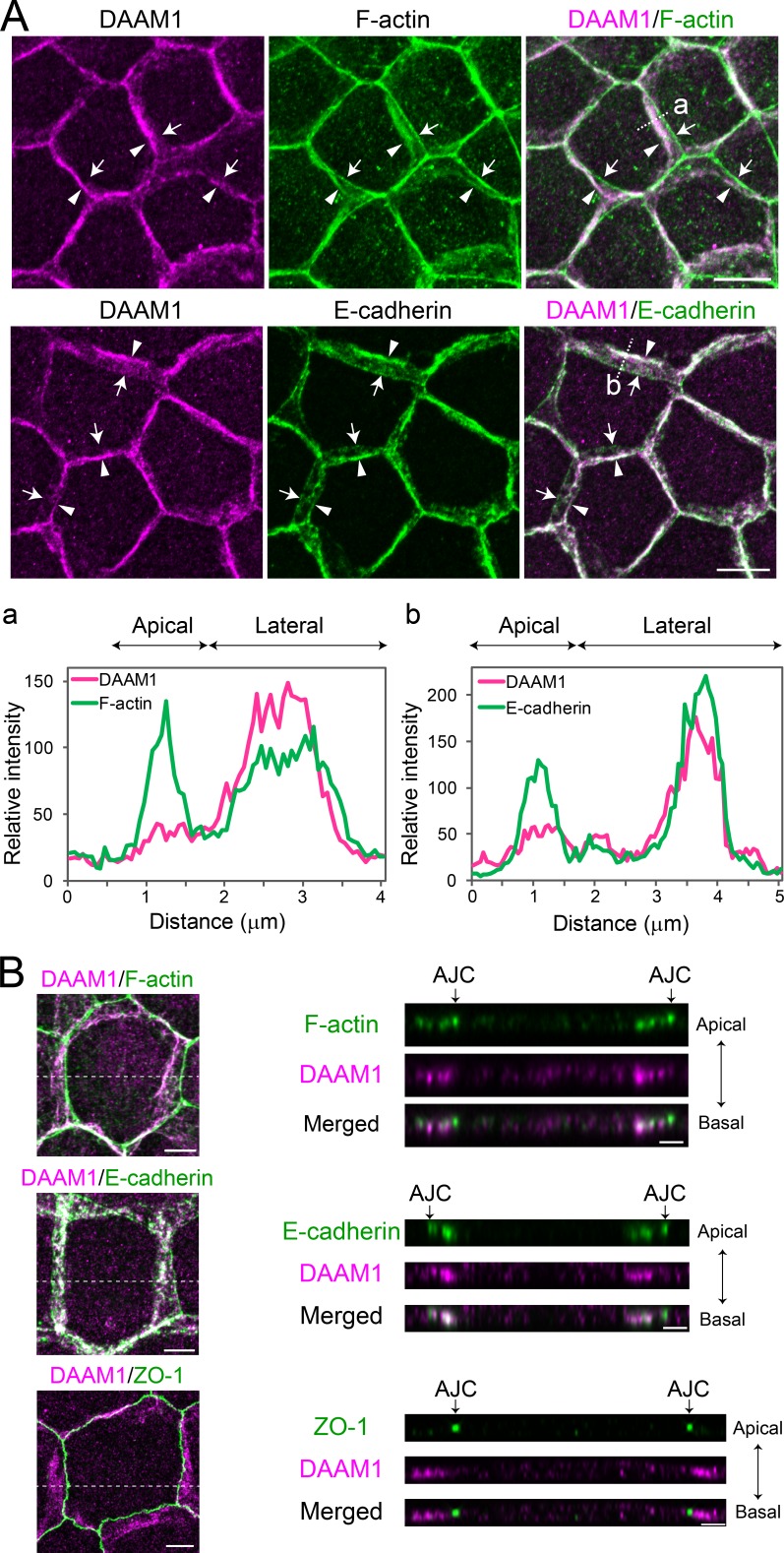
Figure 1.
DAAM1 localizes at the lateral contacts in EpH4 cells. (A) Cells were co-stained for DAAM1 and F-actin (top) or E-cadherin (bottom). Z-stack images are shown. Arrows and arrowheads indicate the apical and basal edge, respectively, of cell junctions. Densitometric traces along the dotted lines (a and b) are also shown. Tracing starts from the apical side. (B) Top (left) and lateral (right) views of cells immunostained for the indicated molecules. Z-stacked confocal images were subjected to super-resolution mode processing. The lateral views were taken along the dashed lines. Arrows indicate AJC positions. Bars: (A) 10 µm; (B, top view) 5 µm; (B, lateral view) 2 µm.

