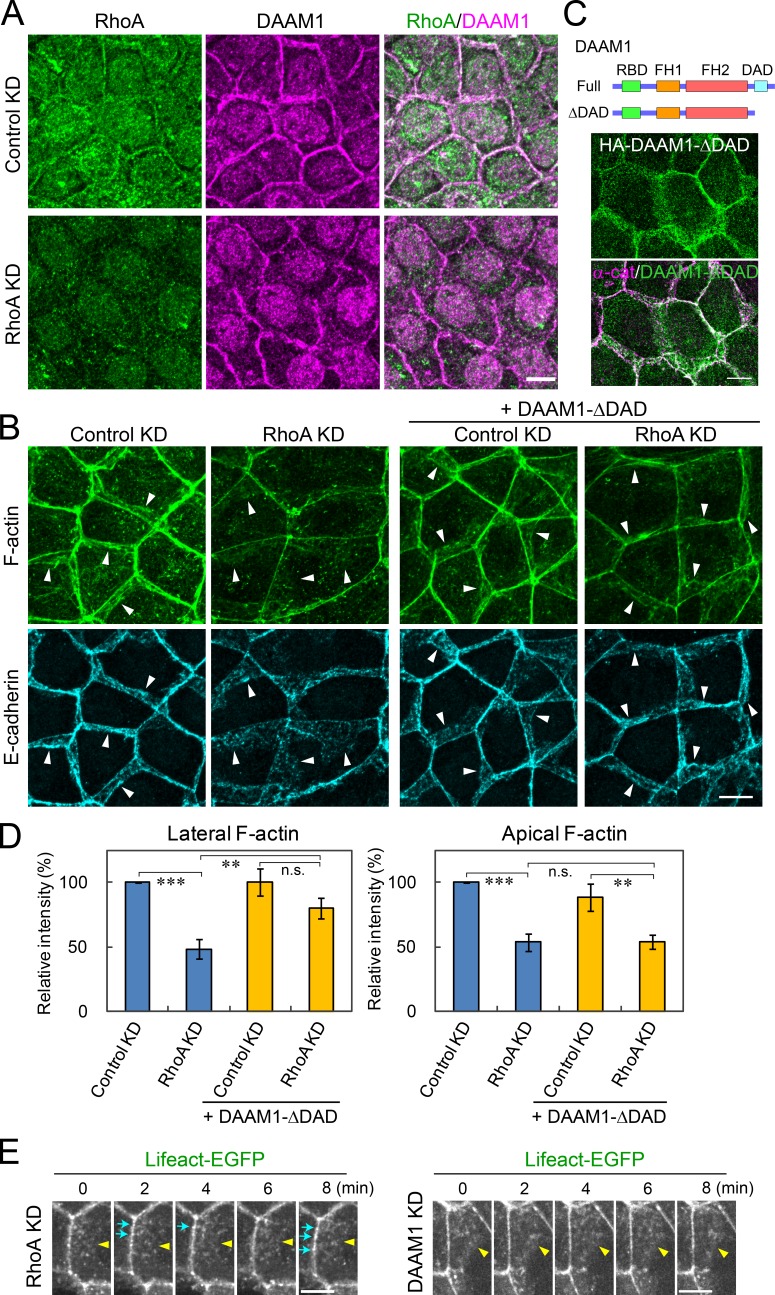
Figure 9.
RhoA regulates cell junctions upstream of DAAM1. (A) Co-immunostaining for RhoA and DAAM1 in EpH4 cells treated with control or RhoA siRNA. Nuclear signals are caused by nonspecific reaction of the primary antibodies. (B) Effects of RhoA depletion and DAAM1-ΔDAD (shown in C) expression on junctional integrity. EpH4 cells or EpH4 cells stably expressing DAAM1-ΔDAD were treated with control or RhoA-specific siRNAs and co-stained for F-actin or E-cadherin. RhoA KD down-regulated not only the lateral but also apical F-actin, and DAAM1-ΔDAD expression restored a nearly normal level of lateral F-actin but not that of apical F-actin in Rho-depleted cells. Arrowheads point to the basal edges of the junctions. (C) The constitutively active DAAM1 mutant DAAM1-ΔDAD. (Top) Schematic representation of the mutant construct. (Bottom) Distribution of HA-tagged DAAM1-ΔDAD in EpH4 cells. α-cat, α-catenin. (D) Quantification of lateral and apical F-actin intensity in the experiments shown in B. Histograms represent the mean of three experiments. Error bars indicate SD. **, P < 0.01; ***, P < 0.001; n.s., not significant. (E) Still images of Video 9, in which Lifeact-EGFP–expressing cells were transfected with RhoA siRNA (left). The distribution of Lifeact-EGFP signals at the AJC changed dynamically. Blue arrows indicate examples of Lifeact-EGFP clusters that were only transiently detectable. Yellow arrowheads point to the basal edges of the junctions. For comparison, still images of Video 2, in which Lifeact-EGFP–expressing cells were transfected with DAAM1 siRNA, are also shown (right). Lifeact-EGFP signals were more stable in DAAM1-depleted than in RhoA-depleted cells at the AJC. (A–C and E) Bars, 10 µm.

