Figure 8.
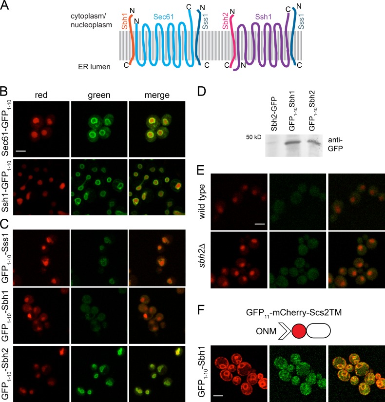
Sbh2, but not Sbh1, localizes to the INM. (A) Schematic of the Sec61 and Ssh1 heterotrimers, including protein topology. (B and C) Localization of Sec61-GFP1–10 and Ssh1-GFP1–10 (B) and N-terminal GFP1–10 fusions to Sss1, Sbh1, and Sbh2 (C). (D) Western blotting with anti-GFP antibodies of the indicated strains shows that GFP1–10-Sbh1 is expressed. The 50-kD molecular mass marker is shown. (E) Localization of GFP1–10-Sbh1 expressed under its native promoter in wild-type and sbh2Δ cells. In B–E, cells contained GFP11-mCherry-Pus1 (red); INM localization based on GFP1–10 (green). (F) GFP1–10-Sbh1 localization at the ONM/ER was tested using the ONM/ER reporter, GFP11-mCherry-Scs2TM. Bars, 2 µm.

