Figure 2.
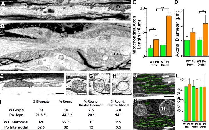
Mitochondrial pathology and MT distribution in juxtaparanodal regions of 1-mo-old P0-CNS optic nerve axons. (A) An electron micrograph shows a mitochondrion in WT juxtaparnodal axoplasm. (B) Mitochondria accumulated in P0-CNS juxtaparanodal axoplasm and were positioned beneath the axolemma. (C and D) Mitochondrial numbers (C) and axonal diameters (D) were similar in WT proximal and distal juxtaparanodal axoplasm (A). Compared with WT axons, mitochondrial numbers (C) were increased in P0-CNS juxtaparanodal axoplasm, and this resulted in increased axonal diameters (D). These increases were greater in distal compared with proximal juxtaparanodal axoplasm (B–D). (E–H) Mitochondrial morphology was binned into elongated with normal cristae (E), rounded with normal cristae (F), rounded with reduced cristae (G), and round with sparse or no cristae (H). (I) Most WT and internodal P0-CNS mitochondria were elongated, but most P0-CNS juxtaparanodal mitochondria were rounded with increased frequency of disrupted cristae. (J and K) MTs were traced in WT (J, green overlay) and P0-CNS juxtaparanodal axoplasm (K, green overlay). Bars, 0.5 µm. (L) MT density was similar in WT and the core of P0-CNS juxtaparanodal axoplasm. MTs were rarely detected in the swollen axoplasm that contained displaced mitochondria (B and K). m, mitochondria. (C, D, and L) Mean ± SEM of axonal values from three animals; *, P < 0.05; **, P < 0.02; t test.

