Figure 6.
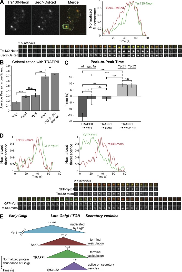
TRAPPII is recruited to the late Golgi immediately after Sec7, coinciding with the Ypt1 to Ypt31/32 transition. (A, bottom) Time-lapse imaging series (2-s intervals) of an example single Golgi compartment (the green boxed region in the Merge panel) in live cells harboring endogenous Trs130-mNeonGreen and Sec7-6xDsRed tags. (top right) Normalized quantification of the Trs130-mNeonGreen and Sec7-6xDsRed signals in the boxed region. (B) Colocalization analysis of TRAPPII (Trs130-mNeonGreen or Trs130-3xmRFPmars) with the early Golgi proteins GFP-Vrg4 and Gea1-3xmRFPmars, the medial/late Golgi localized Rab GFP-Ypt6, the late Golgi/TGN marker Sec7-6xDsRed, and GFP-hFAPP1 PH domain, a coincidence detector of PI(4)P and Arf1. Error bars represent 95% CIs of n ≥ 24 cells. (C) Quantification of peak-to-peak times for each indicated pair of proteins. Error bars represent 95% CIs for n ≥ 10 series. (D) Time-lapse imaging series (2-s intervals) and normalized quantification of GFP-Ypt31 or GFP-Ypt1 and Trs130-3xmRFPmars for a single Golgi compartment as in A. (E) Model for the dynamics of TRAPPII relative to Ypt1, Sec7, and Ypt31/32 at the late Golgi. t = 0 is set to peak TRAPPII recruitment. Regions of interest for time-lapse imaging series are 0.7 × 0.7 µm. Bar, 2 µm. n.s., not significant; **, P < 0.01; ***, P < 0.001.

