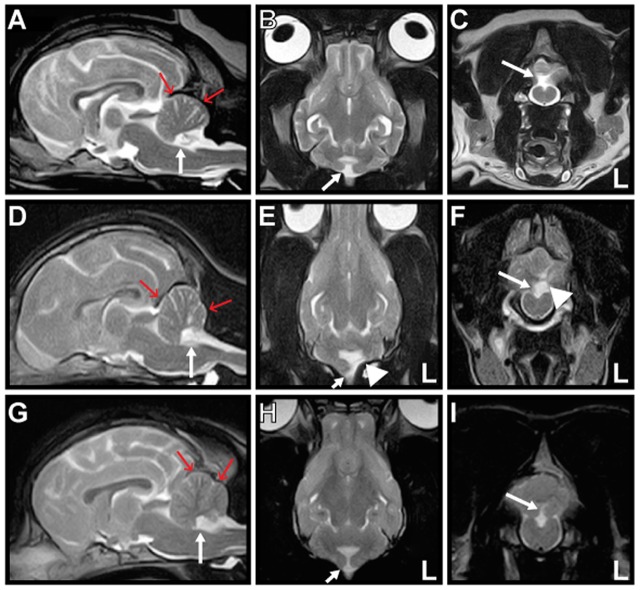
Figure 1.
Magnetic resonance T2-weighted images of Cases 1–3. Mid-sagittal (A,D,G), dorsal (B,E,H), and transverse (C,F,I) images are shown. The nodulus and ventral uvula of the cerebellum (arrow) are absent in all cases. The caudal fossa (red arrows) is reduced in size, indicating hypoplasia in all cases. There is also partial absence of the left cerebellar hemisphere (arrowhead) in Case 2 (E,F).

