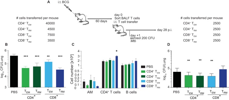
FIG 6 .
Mucosal transfer of airway-resident T cell populations confers protection against TB. (A) B6 mice were BCG vaccinated i.t., and BALF T cell subsets were sorted 60 days later by fluorescence-activated cell sorting. BALF CD4+ and CD8+ TEM and TRM cells were sorted as depicted in Fig. 4A. The sorted T cell population purity was assessed as >86%. From 0.25 × 10⁴ to 4 × 10⁴ sorted cells were i.t. transferred into naive B6 mice (B, D). The numbers of cells transferred are indicated at the top. The following day, recipient mice were aerosol infected with M. tuberculosis and lung CFU counts were determined 28 days later. (B, D) Bacterial CFU counts in lung tissue after BALF removal (B) and in airways and tissue without lavage (D). (C) Immune cellular composition in the lung parenchyma 28 days p.i following i.t. transfer of sorted BALF T cell populations from i.t. BCG-vaccinated mice. Cell numbers are representative of one of two experiments performed as described for panel B. The statistical significance of differences from the i.t. PBS control is shown. Results are presented as mean pooled data ± the standard error of the mean (B to D) from one representative (B, C) or two pooled independent experiments (D) (n = 3 mice per group [B, C] or n = 6 to 8 mice per group [D]). ****, P ≤ 0.0001; ***, P ≤ 0.001; **, P ≤ 0.01; *, P ≤ 0.05. (B to D) Analysis of variance with Tukey’s posttest for significance.

