
FIG 1 .
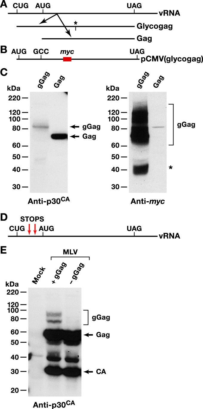
Glycogag expression plasmid and Glycogag-negative MLV clone. (A) Schematic of translation of glycogag and Gag from the viral genome. *, cleavage site in glycogag. Precise location of cleavage site is not known. (B) Schematic of pCMV(glycogag), the glycogag expression plasmid. (C) Lysates of cells transfected with pCMV(glycogag) or Gag expression plasmids were probed with anti-p30CA or anti-Myc antibodies. gGag and Gag bands are indicated. *, cleavage product of gGag. (D) Schematic of glycogag-negative Moloney MLV clone. (E) Lysates of cells transfected with wild-type (+gGag) or glycogag-negative (−gGag) Moloney MLV clones or of mock-transfected cells were probed with anti-p30CA antibody at 48 h posttransfection. gGag, Gag, and capsid (CA) bands are indicated.