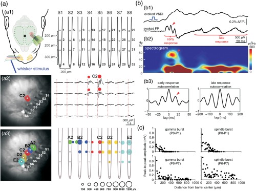
Fig. 2.
Combined VSDI and large-scale multielectrode recordings reveal spatiotemporal properties of sensory-evoked activity in newborn rat barrel cortex in vivo. (a1) Experimental setup combining mechanical single-whisker stimulation, VSDI, and cortical 32-channel recordings using an 8-shank electrode (S1–S8, spacing ). (a2) Selective stimulation of whisker C2 in a P1 rat elicits a local VSD (left) and local electrophysiological (right) responses in the cortical C2 barrel. Note oscillatory response pattern restricted to the upper cortical network with a horizontal extent of . (a3) Color-coded map of the evoked VSD (left) and electrophysiological (right) responses to stimulation of single-whisker A2–E2. Size of the color-coded circle depicts the peak amplitude of electrophysiological response. (b1) Simultaneously recorded VSD and LFP responses in cortical E2 barrel upon stimulation whisker E2 in a P1 rat. Note fast early (gamma burst) and slower late oscillatory responses (spindle burst). (b2) Spectrogram of the LFP response and (b3) autocorrelograms of early and late LFP responses reveals the frequency of the network oscillations. (c) Relationship between LFP amplitude and distance from the barrel center for sensory-evoked gamma bursts (left) and spindle bursts (right) in P0–P1 (upper panels, recordings in four animals) and P6–P7 rats (lower panels, recordings in five animals). Reproduced with permission from Ref. 12.

