Fig. 5.

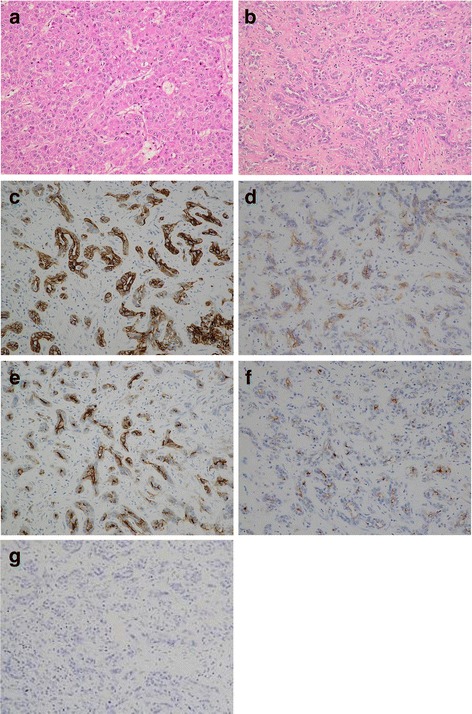
The histopathological examination of the two tumors. a The tumor of the 6th segment of the liver was moderately differentiated hepatocellular carcinoma (Hematoxylin and eosin staining, ×200). b The tumor of the 7th segment of the liver was cholangiolocellular carcinoma (Hematoxylin and eosin staining, ×200). c The immunohistochemical examination of tumor cells from the 7th segment of the liver revealed that they were CK7 positive (×200). d The immunohistochemical examination of tumor cells from the 7th segment of the liver revealed that they were CK19 positive (×200). e The immunohistochemical examination of tumor cells from the 7th segment of the liver revealed that they were EMA positive (×200). f The immunohistochemical examination of tumor cells from the 7th segment of the liver revealed that they were MUC1/DF3 positive (×200). g The immunohistochemical examination of tumor cells from the 7th segment of the liver revealed that they were EpCAM negative (×200)
