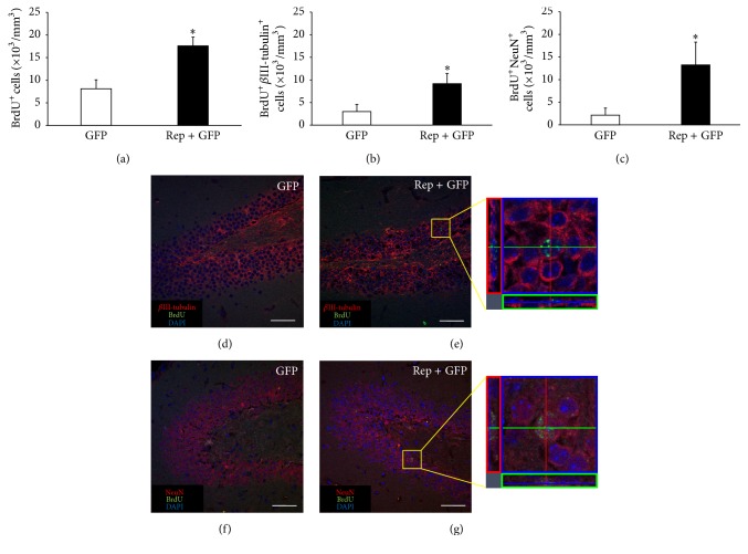
Figure 3.
The number of new neurons in the hippocampus increased by in vivo expression of reprogramming factors. At postnatal 6 weeks, mice were injected with viral vector expressed GFP only (control group) or the four reprogramming factors and GFP (treatment group). To identify newly generated cells, mice were injected daily with 5-bromo-2-deoxyuridine (BrdU) up to 12 days. Eight weeks after injection, histological evaluations were performed. (a) The density of BrdU+ cells in the hippocampus was significantly higher in the treatment group than in the control group (t = 3.528, p < 0.05). (b-c) The density of newly generated neurons was determined through confocal microscopy by calculating the density of cells triple positive for DAPI (blue, nuclei), BrdU (green), and cell type-specific markers such as βIII-tubulin and NeuN. The densities of BrdU+ βIII-tubulin+ (b) and BrdU+NeuN+ (c) cells were significantly higher in the treatment group than the control group (t = 2.450, p < 0.05 and t = 2.297, p < 0.05, resp.). (d–g) Confocal microscope images are immunohistochemistry results. (e, g) Cells with triple positive for DAPI, BrdU, and cell type-specific markers are indicated in the yellow box at the right panel. Scale bars = 50 μm. ∗ p < 0.05.

