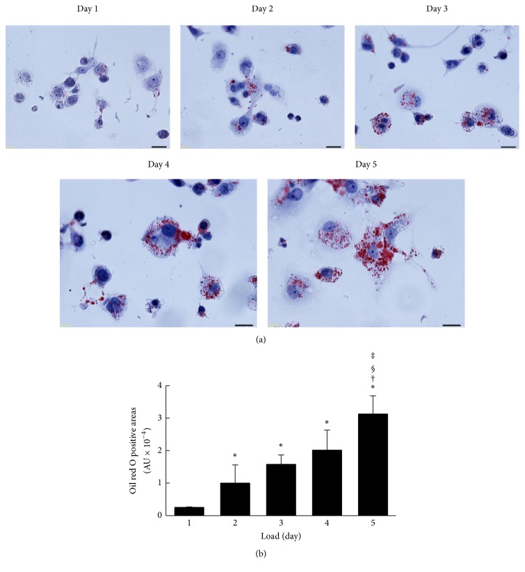
Figure 4.
Foam cell formation. (a) After PMA treatment for 2 days, cells were loaded with acLDL (50 μg protein/mL) and T0901317 (1 μmol/L) for indicated periods. After equilibration, cells were stained with oil red O and hematoxylin (scale bars, 20 μm). (b) Semiquantitative analysis of oil red O positive cells was performed using the ImageJ (National Institutes of Health). The values were indicated by mean + SD (n = 3, ∗ P < 0.05 versus day 1, † P < 0.05 versus day 2, § P < 0.05 versus day 3, and ‡ P < 0.05 versus day 4).

