Figure 6.
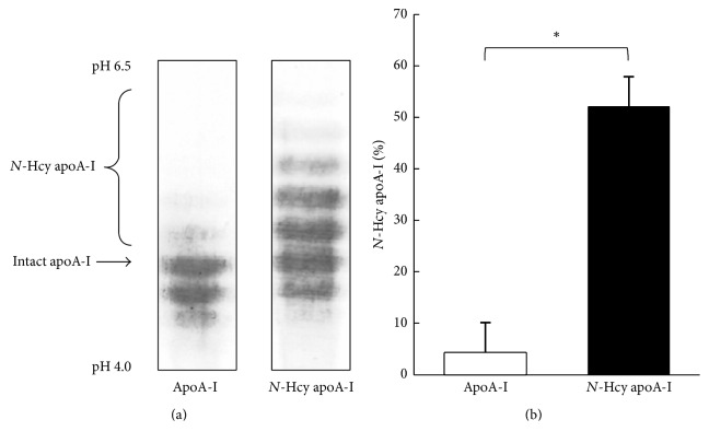
Isoelectric focusing for apoA-I and apoA-I treated with homocysteine thiolactone. (a) Representative isoelectric focusing patterns of apoA-I and N-Hcy apoA-I. Purified apoA-I was incubated with or without 10 mmol/L of homocysteine thiolactone at 37°C for 6 hours. After dialysis against PBS, the samples were analyzed by isoelectric focusing followed by western blotting using anti-apoA-I antibody. (b) Histogram represents the percentage of N-Hcy apoA-I to total apoA-I using CS analyzer. The values were indicated by mean + SD (n = 3, ∗ P < 0.05).

