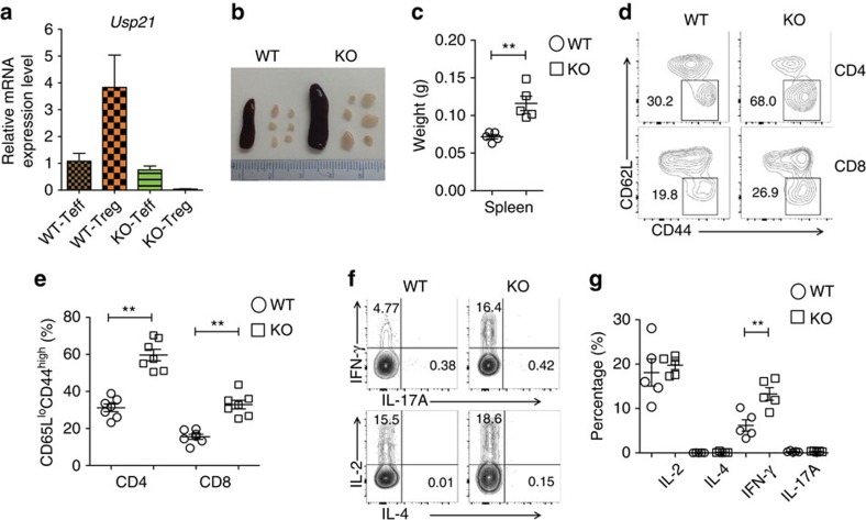
Figure 1. Usp21fl/flFoxp3Cre mice develop spontaneous lymphoproliferative disease.
(a) CD4+CD25−YFP− effector T (Teff) cells and CD4+CD25hiYFP+ Treg cells were sorted from Foxp3Cre (WT) and Usp21fl/flFoxp3Cre (KO) mice. mRNA expression of Usp21 in each population was assessed by qRT–PCR. (n=3 for each group). (b) Image of spleens and peripheral lymph nodes (pLNs) from 8-month-old WT and KO mice. (c) Quantitative analysis of the weight of spleens isolated from WT (n=5) and KO (n=5) mice. (d) Representative figure shown the expression of CD62L and CD44 in splenic CD4+YFP− and CD8+ T cells from WT (n=7) and KO (n=7) littermates. (e) Percentage of splenic CD62LloCD44hi effector memory T cells as in d. (f) Representative figure shown the expression of IFN-γ, IL-17, IL-2 and IL-4 by splenic CD4+YFP− effector T (Teff) cells from WT (n=5) and KO (n=5) mice. (g) Percentage of IFN-γ+, IL-17+, IL-2+ and IL-4+ splenic T cells as in f. Small horizontal lines indicate the mean (±s.d.). All data represent means±s.d. **P≤0.01, as determined by Student's t-test.

