
Figure 5.
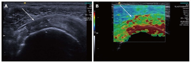
Longitudinal shear wave elastography of a tendinopathic supraspinatus tendon in a different patient to Figure 4. The B mode image (A) shows a partial thickness tear involving the bursal surface fibres (arrow). The corresponding velocity elastogram (B) shows a disorganised pattern with the tear less well delineated (arrow) compared with the B mode image.