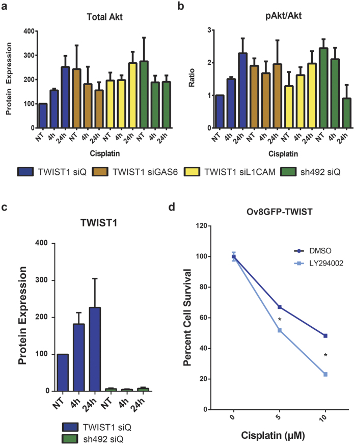
Figure 4. TWIST1, GAS6, and L1CAM expression lead to upregulation of Akt signalling following cisplatin treatment.
(a) Quantification of western blot data shows that over the course of 24 hr, 5 μM cisplatin treatment leads to increased levels of Akt in Ov8GFP-TWIST1 cells (blue), but not in Ov8GFP-sh492 cells (green). Knockdown of GAS6 (brown) or L1CAM (yellow) in Ov8GFP-TWIST1 cells partially abrogates the increase in Akt. NT, not treated with cisplatin. (b) Western blot also reveals an increase in activation of Akt via phosphorylation at Ser 473 over 24 hr of 5 μM cisplatin in TWIST1 expressing cells (blue). The opposite is true in Ov8GFP-sh492 cells, in which Akt activity is reduced over the same time period (green). Knockdown of GAS6 in Ov8GFP-TWIST1 cells (brown) maintains a constant pAkt/Akt ratio, while L1CAM knockdown (yellow) partially prevents Akt activation. (c) Ov8GFP-TWIST1 cells further increase their TWIST1 expression up to 2.3 fold over 24 hr of exposure to 5 μM cisplatin (p=0.0827), whereas Ov8GFP-sh492 cells show no increase. (d) Treatment of Ov8GFP-TWIST1 cells with the PI3K inhibitor LY294002 to prevent Akt activation sensitized cells to cisplatin, compared to DMSO only control, supporting the assertion that Akt signalling is central to TWIST1-driven cisplatin resistance. p < 0.0001 for both concentrations. Error bars represent standard error of the mean.

