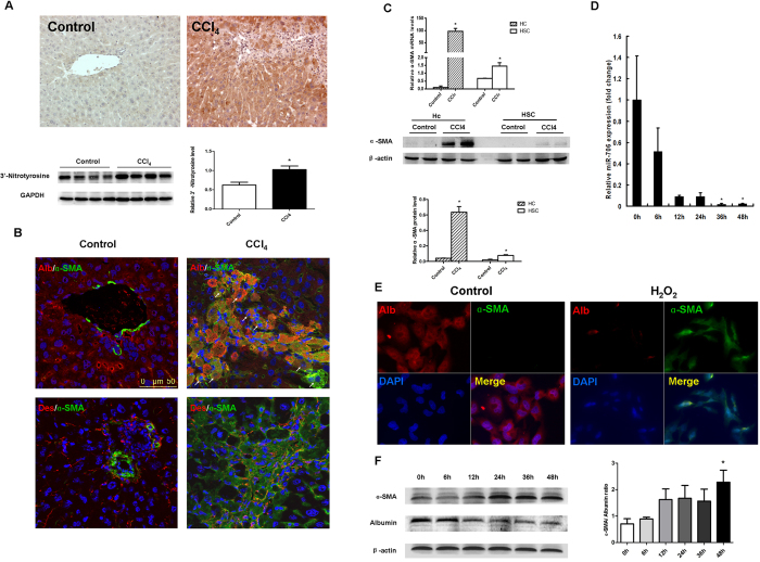
Figure 3. Increased oxidative stress and hepatocyte EMT is characteristic of 4-week CCl4-treated liver.
In vitro treatment with H2O2 induced human hepatocyte cell line L02 EMT, associated with decreased miR-706. (A) Protein levels of 3′-nitrotyrosine in CCl4-treated and control livers were examined by immunohistochemistry and Western blot, *P < 0.05, n = 4 in each group. (B) Representative dual-immunofluorescence staining of albumin and α-SMA, and desmin and α-SMA. (C) mRNA and protein expression for α-SMA in hepatocytes and HSCs isolated from CCl4-treated and control livers. For real-time PCR, GAPDH was used as an internal control. Hc: hepatocytes, *P < 0.05, n = 4 in each group. Data are expressed as mean ± SEM. (D,F) Human hepatocyte cell line L02 was treated with 300 μM H2O2 for 0, 6, 12, 24, 36 and 48 h, respectively. miR-706 expression was examined by RT-PCR (D), *P < 0.05. (E) Protein levels of albumin and α-SMA expression was determined by dual-immunofluorescence staining in 2-day H2O2-treated and control L02 cells, and (F) by Western blot. β–actin was used as loading control.

