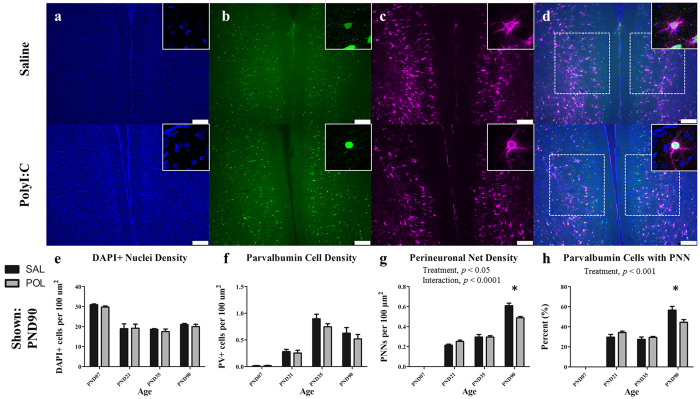
Figure 2. The medial prelimbic cortex has deficits in PNNs that emerge during early adulthood (PND90).
Panels show DAPI (a), PV+ cells (b), PNNs ((c); stained with WFA) from representative rats at PND90. Across both conditions the total number of DAPI labelled nuclei decreased from PND7 to PND21 before plateauing (main effect of Age, p < 0.0001). PV cell density increased from PND7 to PND35 before declining at PND90 (main effect of Age, p < 0.0001). PNN density increased throughout postnatal development with the greatest increases occurring from PND7 to 21, and PND35 to 90 (main effect of Age, p < 0.0001; Age × Treatment, p < 0.0001; Treatment, p < 0.05). In the PND90 cohort, a significant deficit in PNN density emerged in polyI:C treated animals as compared to saline-treated (p < 0.0001). There was also a significant reduction in the number of PV cells ensheathed in a PNN (main effect of Age, p < 0.0001; Treatment, p < 0.001). Insets are representative images from each condition. Scale bar represents 250 μm. *p<0.001.

