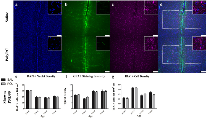
Figure 5. The medial prelimbic cortex exhibited no overt signs of microglial or astrocytic reactivity (PND90 shown).
Panels show DAPI (a), IBA1 (c) and GFAP (b). Across both conditions the total number of DAPI labelled nuclei decreased throughout postnatal development (main effect of Age, p < 0.0001). Microglial density was increased slightly throughout postnatal development aside from a dramatic spike in density in the PND21 cohort (main effect of Age, p < 0.001; Age × Treatment, p = 0.056). GFAP staining was sparsely present within the cortical parenchyma, instead clustering around the cortical surface and blood vessels throughout the tissue. GFAP staining intensity increased from PND21 to PND35 where it leveled off into maturity (main effect of Age, p < 0.0001). Neither polyI:C treated animals or controls showed significant differences between either of these immune markers. Insets are representative images from each condition. Scale bar represents 250 μm. *p < 0.05.

