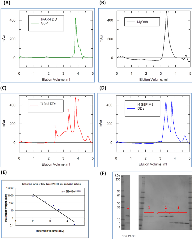
Figure 3. Phosphorylated Ser8 IRAK-4 death domain interferes with the Myddosome formation.
(A–D) Absorbance Elution spectrum of a TSKgel SuperSW3000 analytical size exclusion column at 280 nm. (A) 0.5 mg.ml−1 of IRAK-4 death domain only. (B) 0.5 mg.ml−1 of MyD88 DD only. (C) 1:1 mix of MyD88 and non-phosphorylated IRAK-4 death domains concentrated to 0.5 mg.ml−1 prior to loading. (D) 1:1 mix of MyD88 and Ser8 phosphorylated IRAK-4 death domains concentrated to 0.5 mg.ml−1 prior loading. (E) Calibration of gel filtration using protein standard markers (F) 4–20% reducing Tris-Glycine SDS PAGE of fractions corresponding to peak 1, 2 and 3 of figure 15a. Fractions corresponding to peak 1 were concentrated using a 3 kDa MW cut-off Amicon Spin Filters for better visualisation of samples. One of three repeats is shown.

