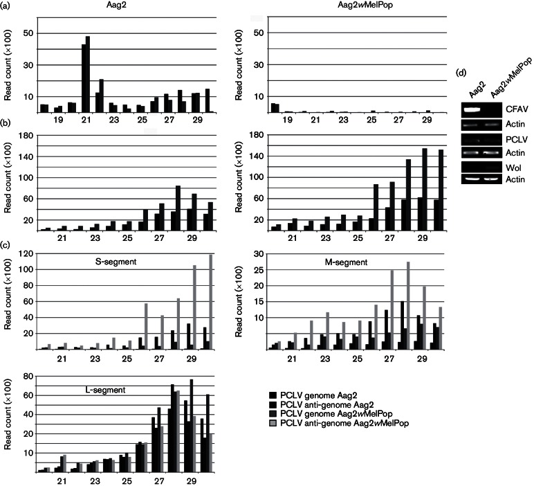
Fig. 1.
Presence or absence of CFAV, PCLV and wMelPop in Aag2 and Aag2wMelPop cells. Size distribution of small RNA molecules mapping to the CFAV (a) or PCLV (b) genome (black)/anti-genome (grey) in A. aegypti-derived Aag2 or wMelPop-transinfected Aag2 cells. (c) Size distribution of small RNA molecules mapping to the different segments of PCLV (S, M and L) genome/anti-genome in A. aegypti-derived Aag2 or wMelPop-transinfected Aag2 cells. (d) Detection of CFAV or PCLV in Aag2 and Aag2wMelPop cells by RT-PCR. Actin was used as loading control.

