
Fig. 1.
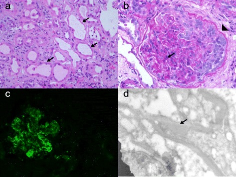
Microscopic examinations of kidney biopsy specimens. a and b Hematoxylin and eosin staining of the kidney biopsy specimen revealed interstitial infiltrate with tubular injury (arrows), glomerulonephritis with cellular crescents (arrowhead), and mesangial proliferation (arrow). c Immunofluorescence staining for IgA deposits. d Electron microscopic picture of subepithelial deposit (arrow)