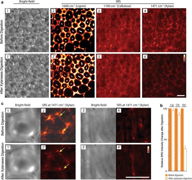
Fig. 6.

In situ tracking of lignin, cellulose, and xylan in deacetylated corn stover cell walls before and after xylan digestion by SRS. a Comparison of bright-field images of cell walls and SRS images of lignin, cellulose and xylan in the same cell walls. b Comparison of the relative overall SRS signal change in the images (before enzyme digestion = 100%) (Lig lignin, Cel cellulose and Xyl xylan; error bar are from 5 repeat digestion experiments). Lignin and cellulose are not affected by xylanases, and xylan is significantly reduced due to xylanases digestion. c Zoom-in bright-field images of cell wall and SRS images of xylan in two areas in vascular bundle region (c 1–4: before xylan digestion; and c 1′–4′: after xylan digestion) show significant xylan distribution changes in the cell wall due to the heterogeneous enzymatic digestion. Raman frequencies used for SRS imaging: lignin—1600 cm−1, cellulose—1100 cm−1 and xylan—1471 cm−1. Scale bar = 20 µm
