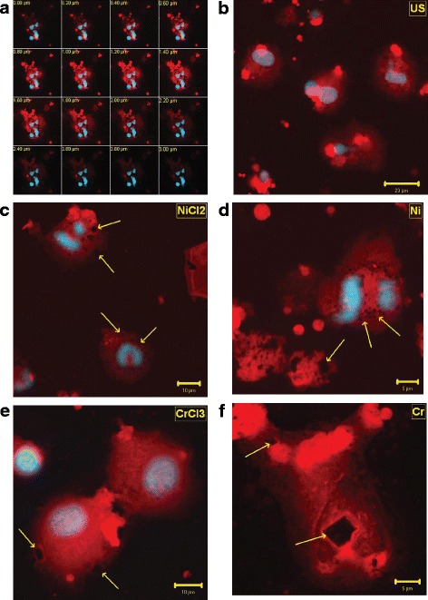
Fig. 4.

a-f Confocal Microscopy. Representative images of z-stacks optical sections (a), untreated cells (b), NiCl2 (c), Ni (d), CrCl3 (e), and Cr (f) -stimulated cells. (Phalloidin TRITC-conjugate, Hoechst 33258). (Phalloidin TRITC-conjugate, Hoechst 33258). b = 40x, c,e = 63x, d,f = 100x original magnification
