Fig. 2.

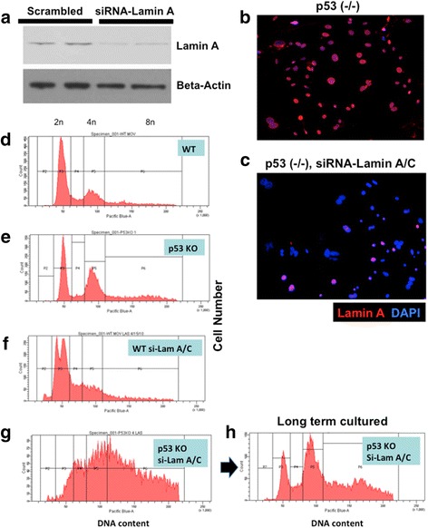
Lamin A/C suppression in primary mouse ovarian surface epithelial (MOSE) cells results in aneuploidy and polyploidy, synergistic with p53 deletion. Primary wildtype (WT) and p53 knockout (KO) MOSE cells were transfected with control (scrambled) or siRNA (si-Lam A) to suppress lamin A/C suppression. a At day 3, the lamin A/C-suppressed cells were analyzed by Western blot for the presence of lamin A protein. Duplicate experiments are shown for control (scrambled siRNA) and siRNA specific to mouse lamin A/C. b The cells were analyzed by immunofluorescence microscopy for the expression of lamin A. An example of p53 (-/-) MOSE cells treated with control siRNA is shown. c In comparison, staining was reduced in cells treated with siRNA-lamin A/C. d Flow cytometry was performed 3 days after siRNA transfection. Cells were collected following trypsin digestion, washed with PBS, and cell pellets were resuspended in ice cold ethanol/PBS (70 % v/v) with gentle agitation. The fixed cells were kept at − 20°C until ready to use. Prior to flow cytometric analysis, cells were centrifuged at 1200 rpm for 5 min and washed twice with PBS before resuspension in 0.5 mm vybrant violet dye. Cells were then incubated at 37°C for 30 min before flow cytometric analysis for DNA content. Flow cytometry profile for wildtype (WT) cells treated with control siRNA is shown. e p53 knockout cells; f WT cells treated with siRNA-lamin A/C; g p53 knockout cells treated with siRNA-lamin A/C. h Flow cytometry profile of the p53 knockout, siRNA-lamin A/C-treated MOSE cells following longer-term (2 months) culturing
