Fig. 6.

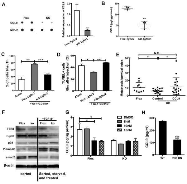
CCL9 is critical in myeloid-specific TGF-β regulation of tumor metastasis. (A) Cytokine protein array indicating CCL9 expression in floxed or TβRII-deficient Gr-1+CD11b+ cells sorted from peripheral blood of 4T1 tumor-bearing Tgfbr2flox or Tgfbr2MyeKO mice. The samples were combined from 3 mice, with duplicates for each sample. Semi-quantitative data of dot density is on the right. (B) CCL9 ELISA in myeloid cells sorted from peripheral blood of TβRII-deficient or control tumor-bearing mice (n=3). (C) SCVM for tumor cell survival. GFP labeled 67NR cells (5×10⁵) were co-injected with floxed or TβRII-deficient Gr-1+CD11b+ cells (1×10⁶). The lungs were taken out for images 1 and 6 hours after injection. Ten fields for each mouse lung were examined. Fluorescent signals at 6 hours were normalized with 1h signal. n=3 mice per group. (D) TUNEL assay for tumor cell apoptosis from lung sections from (C). TUNEL positive cells were counted and calculated and presented as percentage of GFP+ cells as shown at Y-axis. (E) PUMA for tumor cell survival and metastasis. 67NR-GFP cells were co-injected with TβRII-deficient Gr-1+CD11b+ cells with or without CCL9 over-expression. Fluorescence imaging was obtained 14 days after lung section culture. Fluorescence signal per field was quantified then normalized to day 0 signal and presented as metastasis survival index. Three mice each group, 3-4 lung pieces each mouse. (F) Western blot for P-p38 level in TβRII-deficient Gr-1+CD11b+ cells, compared with control cells, with or without TGF-β1 treatment. One representative experiment is shown here from two performed. (G) CCL9 ELISA of floxed or TβRII-deficient Gr-1+CD11b+ cells treated with p38 inhibitor for 6 hours. (H) CCL9 ELISA of Gr-1+CD11b+ cells sorted from spleen of P38 dominant negative or wild type mice. The cells were cultured in 4T1 tumor conditioned media for 24 hours. Spleens from 3 mice were pooled for sorting. Validation of P-p38 by Western is in the lower panel. *P<0.05, **P<0.01, ***P<0.001.
