Fig. 5.

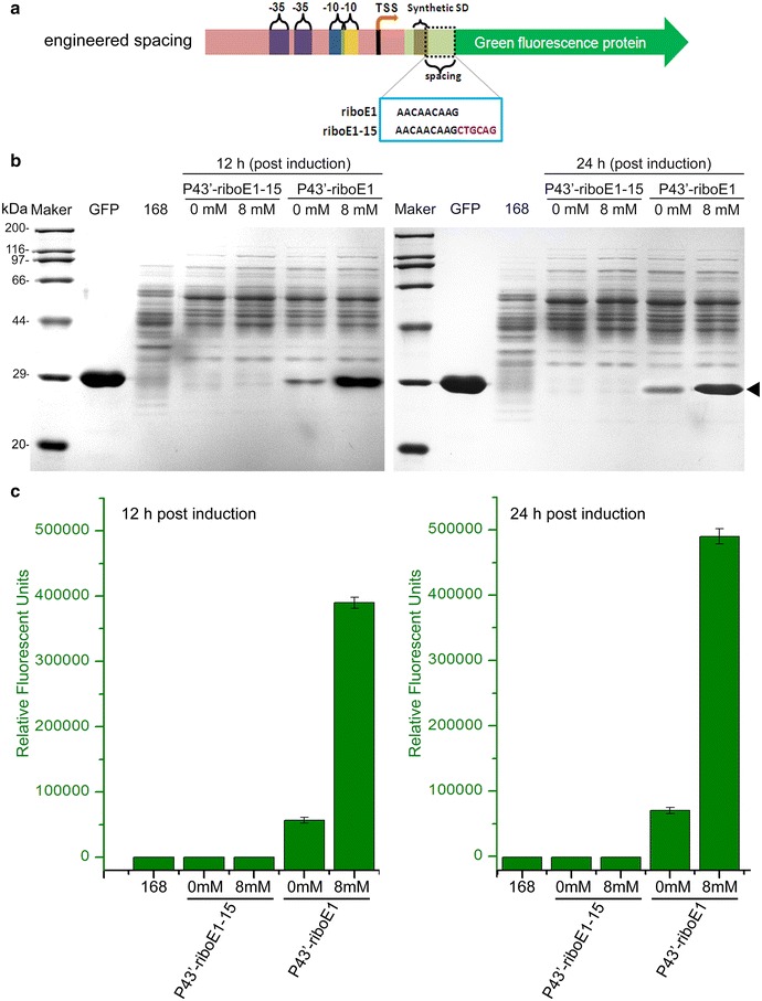
Determination of the effect of length of spacer on the induced level of GFP expression. a Schematic diagram displays the gene structure of riboE1, and riboE1-15 harbouring 15-bp spacer between SD sequence and start codon was produced by insertion of PstI restriction site immediately downstream of the SD sequence. The SD sequences within riboE1 and the modified riboE1-15 are in the azure box. b SDS-PAGE analysis was carried out to detect the expression level of GFP driven by P43′-riboE1 and P43′-riboE1-15 after induction with 8 mM theophylline for both 12 and 24 h. The strains harbouring recombinant plasmids treated with 4% DMSO are designated as 0 mM (controls). Protein extracts from Bacillus subtilis 168, which were collected at 19 and 31-h culture (equal to the induced groups with 12 and 24-h induction, respectively), are used as the negative controls. The GFP stands for the purified GFP protein, which is served as the specific marker to indicate the corresponding position of heterologous GFP. c Measurement of the relative fluorescent units of GFP corresponding to the SDS-PAGE in b
