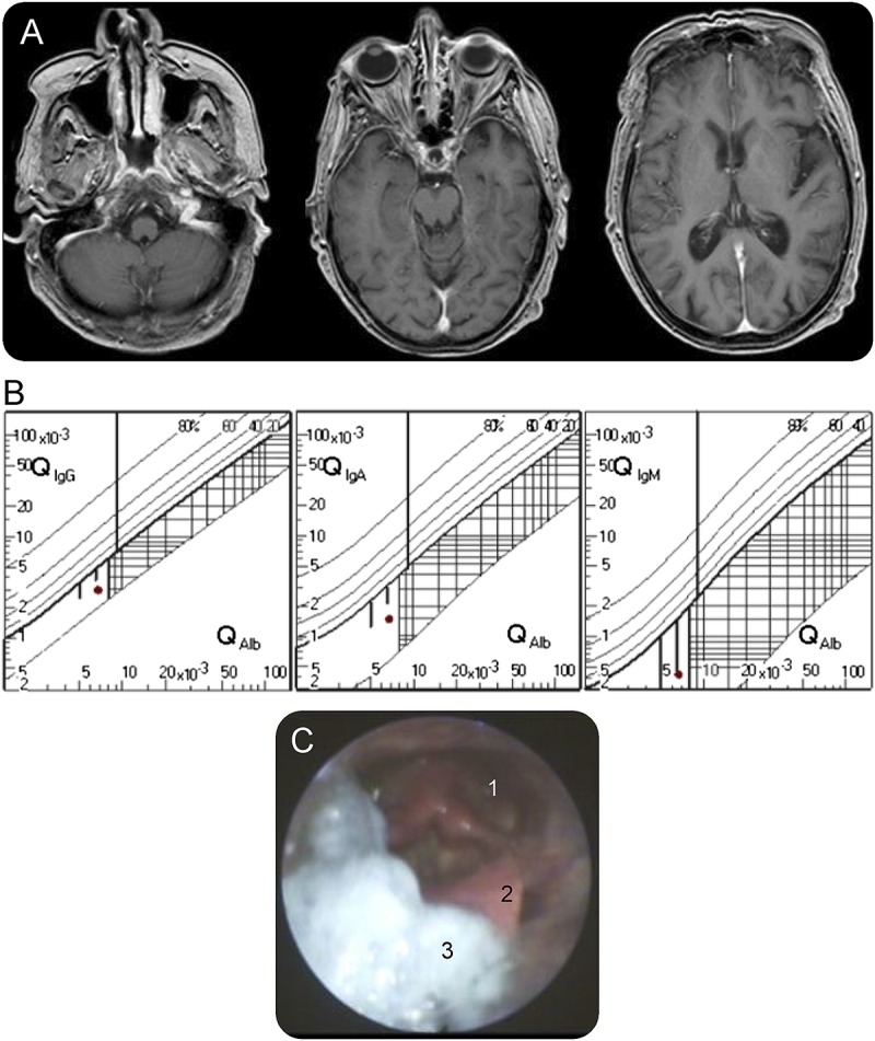
Figure. Investigations.

(A) T1 gadolinium-enhanced axial MRI shows no evidence of underlying brainstem or cranial nerve pathology. (B) No intrathecal immunoglobulin synthesis was observed in CSF analysis using the Reiber scheme. (C) Fiber optic endoscopic evaluation of swallowing reveals postdeglutitive residue in the valleculae epiglotticae (3), on top of the epiglottis (2), and in the piriform sinuses (1).
