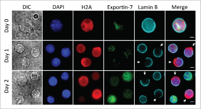
Figure 3.

Localization of exportin-7 in different stages of mouse fetal liver erythropoiesis. Ter119-negative E13.5 mouse fetal liver erythroblasts were purified and cultured for indicated days in an erythropoietin-containing medium. Immunofluorescence stains for exportin-7, H2A, Lamin B and DNA (DAPI) were performed. Arrows indicate nuclear opening. Scale bars: 5 μM.
