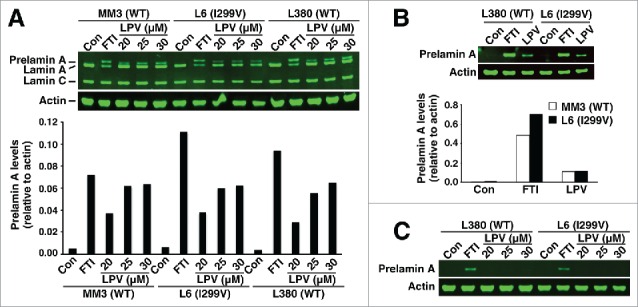
Figure 4.

Western blots of cell extracts from wild-type fibroblasts (MM3 and L380) and I299V fibroblasts (L6). Wild-type and I299V fibroblasts were grown without a drug or in the presence of a protein farnesyltransferase inhibitor (FTI) or lopinavir (LPV). (A) Western blot of fibroblast extracts with the lamin A/C polyclonal antibody. Bar graph shows the amounts of prelamin A, relative to actin, as judged by an Odyssey infrared scanner. (B) Western blot of fibroblast extracts with antibody 3C8. The LPV concentration was 25 μM. Bar graph shows the amounts of prelamin A, relative to actin. (C) Western blot of fibroblast extracts with antibody 7G11.
