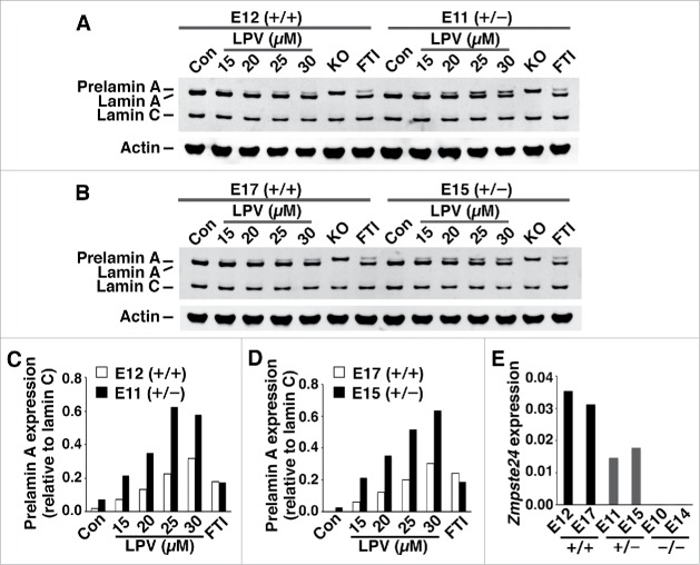
Figure 5.
Prelamin A accumulation in wild-type mouse fibroblasts (Zmpste24+/+) and heterozygous Zmpste24 knockout fibroblasts (Zmpste24+/−) in response to lopinavir treatment. (A–B) Western blots, using a lamin A/C polyclonal antibody, of extracts from Zmpste24+/+ fibroblasts (E12, E17) and Zmpste24+/− fibroblasts (E11, E15) after lopinavir treatment. In these studies, Zmpste24−/− fibroblasts were included as controls (KO). (C–D) Bar graphs show amounts of prelamin A, relative to lamin C, in fibroblasts E12 and E11 (C) and in fibroblasts E17 and E15 (D). (E) Half-normal levels of Zmpste24 transcripts in Zmpste24+/− fibroblasts (E11, E15), compared with Zmpste24+/+ fibroblasts (E12, E17). As controls, we included 2 different Zmpste24−/− fibroblast cell lines (E10, E14).

