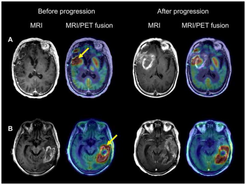
Figure 1. T1-Gad MRI and co-registered MRI/AMT-PET fusion images of two patients with tumor progression beyond the PET+ region.

(A) Patient #1 showed contrast-enhancement and high AMT uptake at baseline in the right inferior frontal and anterior temporal lobes. The temporal lobe area with AMT increase extended well beyond the contrast-enhancing region medially toward the basal ganglia (arrow), with a maximum standardized uptake value (SUVmax) of 5.7. Follow-up MRI in one month showed a rapid progression with a large medial extension of the contrast-enhancing lesion encompassing and extending beyond the baseline PET+ area.
(B) Patient #8 had a large contrast-enhancing area and high AMT uptake in the left temporal region. The PET+ area extended anterior to the Gad+ lesion (arrow), with an SUVmax of 5.4. T1-Gad MRI 5 months later showed extension of the contrast enhancement into the anterior temporal lobe encompassing and extending beyond the baseline PET+ region.
