
Figure 4.
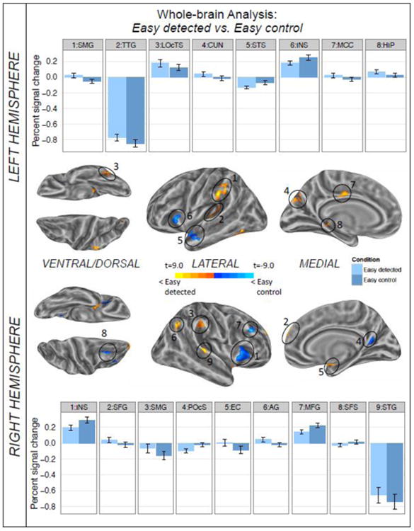
Results of the whole-brain comparison between the easy detected and easy control condition for the time window of 3-12 sec post sentence onset shown on inflated average brain surfaces of the left and right hemisphere. Labels refer to the local activation maximum of each cluster. The individual per-vertex threshold was p < .01 (corrected FWE p < .05). Warm colors indicate higher levels of activation for easy detected; cold colors reflect greater activation for the easy control sentences. Bar graphs present the difference between conditions for the local activation maximum of each cluster. Error bars depict standard error of the mean. AG=angular gyrus, CUN=cuneus, EC=entorhinal cortex, HIP= hippocampus, INS= anterior insula, LOcTS=lateral occipito-temporal sulcus, MCC=middle cingulate cortex, MFG=middle frontal gyrus, POcS=posterior occipital sulcus, SFS=superior frontal sulcus, SFG=superior frontal gyrus, SMG=supramarginal gyrus, STS=superior temporal sulcus, STG=superior temporal gyrus, TTG=transverse temporal gyrus.