
Figure 3.
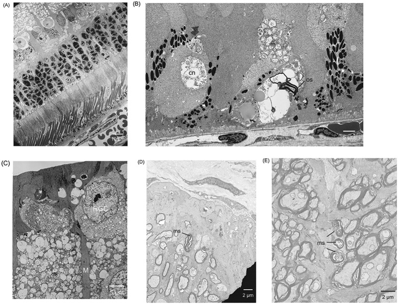
Electron microscopy. A) Rods and cones are clearly delineated in the WT rabbit. (B) Retinal pigment epithelium pigment granules irregularly distributed. Degeneration of cone nuclei, clump of mitochondria, and debris of outer segments are observed in the Tg rabbit. (C) Vacuolar degeneration of retinal ganglion cells (RGCs) and hypertrophic proliferation of Müller cells are observed. (D) Degeneration of myelinated axons in the optic disc. (E) Damage of the ring-like structure of the myelin sheath of small axons in the optic nerve. INL = inner nuclear layer; ONL = outer nuclear layer; R = rod; C = cone; cn = cone nuclei; m = mitochondria; os = outer segment; RGC = retinal ganglion cell; M = Müller cell; ms = myelin sheath.