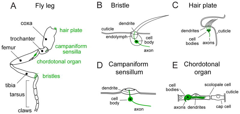
Figure 1. Anatomy of mechanoreceptor organs on the fly leg.
(A) Schematic of the Drosophila leg, illustrating the four classes of mechanoreceptor organs. Note that these examples do not represent the full complement of leg mechanoreceptors, but rather illustrative examples of each type of organ.
(B) Mechanosensory bristles are the primary exteroceptive organs, densely tiling the fly cuticle. Deflection of the bristle leads to firing in the bristle sensory neuron. In other insects, bristles are known as tactile hairs.
(C) Campaniform sensilla are small domes, which detect tension and compression in the surrounding cuticle. They are often found clumped in fields where strains on the cuticle are likely to be high, such as on proximal regions of the leg.
(D) Hair plates are tightly packed groups of small, stiff, parallel hairs, each of which is innervated by a single sensory neuron. They are often positioned next to folds within the cuticle, so that the hairs are deflected during joint movement. They function as proprioceptors, sensing movements of one joint segment relative to the adjoining segment.
(E) Chordotonal organs are stretch-sensitive mechanoreceptors that contain many individual sensory neurons with diverse mechanical sensitivities. They are found at leg joints, where they encode the angle and movement of the leg, as well as Johnston’s organ in the fly antenna, where they encode auditory signals.

