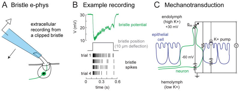
Figure 2. Mechanosensitive properties of tactile hair neurons.
(A) Extracellular electrophysiological recordings from tactile hairs can be made by inserting the shaft of a clipped hair in a recording pipette, which is mounted on a piezoelectric actuator so that the pipette can be used to move the hair.
(B) An example extracellular recording from a fly femur bristle [166] shows a downward field potential deflection (the receptor potential) and a burst of superimposed action potentials. Repeated movement of the bristle leads to adaptation in the bristle response, depicted as a spike raster.
(C) The equivalent circuit of the tactile hair epithelium recording [adapted from 20]. The hair neuron dendrite is bathed in endolymph (high K+), which is separated from the circulating hemolymph (low K+) by an electrically tight layer of epithelial cells. The K+ gradient is established by pumps in the apical membranes of epithelial cells, depicted here as a current source. The transepithelial potential (measured in the endolymph relative to the hemolymph) is about +30 mV. If the neuron rests at about −60 mV, then the total driving force pushing K+ into the neuron’s dendrite through mechanotransduction channels (gm) would be about 90 mV. It should be noted that the tactile hair neuron is separated from the hemolymph by a layer of glial cells (not depicted here). Membrane capacitance is also omitted for clarity.

