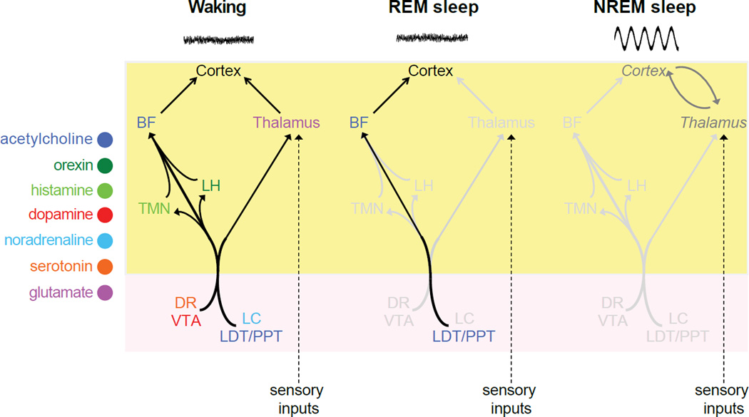
Figure 3. Neuroanatomical pathways by which neurotransmitter systems regulate arousal in the mammalian brain.
The cell bodies of each neurotransmitter system are located in brain regions whose names are colored and abbreviated as follows: BF (basal forebrain), LH (lateral hypothalamus), TMN (tuberomamillary nucleus), DR (dorsal raphe nucleus), VTA (ventral tegmental area), LC (locus coeruleus), LDT/PPT (laterodorsal tegmental and pedunculopontine nuclei). Not shown: GABAergic inhibition of most of these brain regions by the ventrolateral preoptic nucleus to promote sleep. During waking, the cortex is excited by ventral and dorsal pathways through the basal forebrain and thalamus, respectively. During REM sleep, aminergic signaling is reduced, thus blocking sensory throughput at the level of the thalamus, but the persistence of cholinergic signaling through the ventral pathway continues to excite the cortex. During NREM sleep, aminergic and cholinergic signaling are both reduced, leading to lowered cortical activation, the appearance of SWA, and its entrainment by thalamocortical loops. Yellow and pink areas represent the forebrain and brainstem, respectively.

