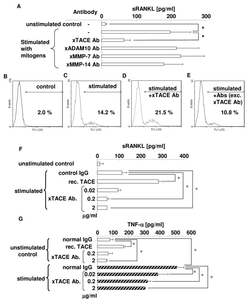
Figure 2. TACE is the most important proteolytic enzyme with RANKL sheddase properties in activated PBLs.
PBLs were stimulated with PMA (50 ng/ml), ionomycin (1 μg/ml), and LPS (10 μg/ml) in the presence or absence of specific neutralizing antibodies for each potential RANKL sheddase, followed by collection of culture supernatant. Subsequently, sRANKL concentrations were measured by ELISA in triplicate wells for each group (Fig. 2A) * p<0.05. mRANKL expression on plasma membrane of PBLs was examined by flow cytometry. Cells on the lymphocyte gate, as defined by forward and side scatter characteristics, were investigated. Representative data of unstimulated PBLs (Fig. 2B), stimulated PBLs (Fig. 2C), PBLs stimulated in the presence of anti-TACE specific antibody (Fig. 2D), and PBLs stimulated in the presence of antibodies, except anti-TACE antibody (combination of anti-ADAM-10 antibody, anti-MMP-7 antibody, and anti-MMP-14 antibody) (Fig. 2E), are shown. Percentages in the figure indicate mRANKL-positive cells in the population (2.0, 14.2, 21.5, and 10.8%, respectively).
In order to confirm the functionality of anti-TACE antibody in neutralizing the TACE activity, PBLs were stimulated with PMA (50 ng/ml), ionomycin (1 μg/ml), and LPS (10 μg/ml) in the presence or absence of recombinant TACE (0.7 μg/ml) or anti-TACE antibody (0.02, 0.2, or 2 μg/ml). The collected culture supernatants were subjected to ELISA for the measurement of sRANKL (Fig. 2F) and TNF-α concentrations (Fig. 2G) in triplicate wells for each group. * p<0.05

